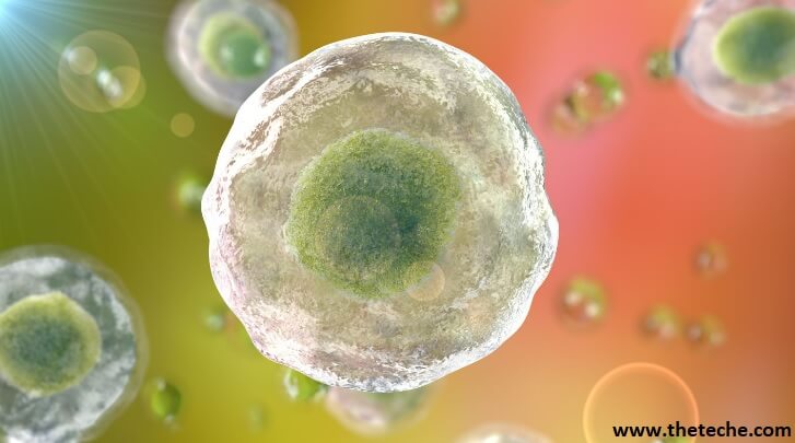
Structure enclosed by the cell membrane is known as cytoplasm, which is aqueous, semitransparent, elastic, and thick. Cytoplasm consists of 80% water and remaining 20% include proteins (enzymes), carbohydrates, lipids, inorganic ions, and many low-molecular weight compounds.
In the cytoplasm, most of the vital chemical reactions (metabolism) of the cells take place.
In prokaryotic cytoplasm, the major structures are nucleoid, ribosomes, and various reserve deposits (called inclusion bodies). Most of the membrane-bound structures, e.g., endoplasmic reticulum, mitochondria, Golgi apparatus, lysosomes, and membrane-bound vacuoles are not present in the prokaryotic cytoplasm.
Nucleoid – Prokaryotic cells lack the presence of a well-developed nucleus or a nucleus with a distinct membrane or a mitotic apparatus or a nucleolus. Yet, their nuclear structure is encompassed in the form of a double stranded DNA (dsDNA) present close to the centre of the cell.
This nuclear structure is labelled as a nucleoid/nucleoplasm region/chromatin body/nuclear equivalent/bacterial chromosome, as it is not a distinct nucleus. The nucleoid can be visualised by a light microscope using Feulgen staining, which is specifically used to visualise DNA.
It gives the impression of a light area with a delicate fibrillar structure under an electron microscope.
The genetic information of a bacterial cell is contained in the nucleoid which carries all the information essential for the structure and function of bacterial cell. It is present in the form of a continuous, long thread of single double-stranded DNA, arranged in a circular manner.
Ribosomes – Ribosomes are very small structures, present in the cytoplasm of a bacterial cell, ranging in number from 15,000-20,000. They are responsible for the characteristic granular appearance of the cytoplasm.
The process of translation (protein synthesis) is carried out by ribosomes. In this process, the message in nucleotides is carried by the messenger RNA (mRNA) from the genome to the ribosome, where proteins are formed by the linking of amino acids through peptide bonds.
Approximately, 40% of the dry weight of a bacterial cell is accounted for the ribosomes. Ribosomes are comprised of ribosomal Ribonucleic Acid (rRNA) and proteins. Some bacterial ribosomes are suspended freely within the cytoplasm, and the remaining ribosomes are linked to the inner surface of the cytoplasmic membrane, and are engaged in protein synthesis (to be ultimately transported out of the cell).
Inclusion Bodies – In the cytoplasm of prokaryotic cells, some reserve deposits, known as inclusions are present. Cells store certain nutrients present in large amounts to utilise them in deficient conditions. It has been proved that macromolecules in inclusions to prevent the increase in osmotic pressure that would result if the molecules were dispersed in the cytoplasm.
Some inclusions are commonly present in wide variety of bacteria, while some are limited to a small number of species, thus aiding in their identification.
Given below are the different inclusion bodies :
Metachromatic Granules : The name of these large inclusions has been derived from the fact that at times they stain red with certain blue dyes (e.g., methylene blue). Collectively they are termed volutin, which is a reserve of inorganic phosphate (polyphosphate) used for ATP synthesis.
It is formed by cells grown in phosphate-rich environments. Algae, fungi, protozoa, and bacteria contain metachromatic granules, which are characteristic of Corynebacterium diphtheriae (diphtheria causing agent), thus are diagnostically significant.
Polysaccharide Granules : These inclusions contain glycogen and starch. The glycogen granules appear reddish brown and starch granules appear blue when bacterial cells are stained with iodine, thus aiding the identification of these inclusions.
Lipid Inclusions : These inclusions are present in various species of Mycobacterium, Bacillus, Azotobacter, Spirillum, etc. A common lipid-storage material (unique to bacteria) is poly- β-hydroxybutyric acid (a polymer). The presence of these inclusions is identified by staining the bacterial cells with fat-soluble dyes, e.g., Sudan dyes
Sulphur Granules : The “sulphur bacteria” (genus Thiobacillus) obtain energy from the oxidation of sulphur and sulphur-containing compounds. These bacteria deposit sulphur granules in the bacterial cell to act as a source of energy.
Carboxysomes : These inclusions contain ribulose 1,5-diphosphate carboxylase enzyme. Bacteria obtaining carbon from carbon dioxide utilise this enzyme for carbon dioxide fixation during photosynthesis. Nitrifying bacteria, cyanobacteria, and thiobacilli contain these inclusions.
Gas Vacuoles : These hollow cavities are present in some aquatic prokaryotes, including cyanobacteria, anoxygenic photosynthetic bacteria, and halobacteria. Each vacuole has rows of several individual gas vesicles (hollow cylinders covered with protein). Gas vacuoles maintain buoyancy so that the bacterial cells remain in the water at a depth where they can receive optimum amounts of oxygen, light, and nutrients.
Magnetosomes : These inclusions of iron oxide (Fe3O4) are formed by some gramnegative bacteria (Aquaspirillum magnetotacticum) and act like magnets. Bacteria with the help of these inclusions move downward to a suitable attachment site.
Magnetosomes decompose hydrogen peroxide formed in the bacterial cells in the presence of oxygen. Researchers have guessed that magnetosomes provide protection to the cells by avoiding accumulation of hydrogen peroxide. Various culture methods are under development by industrial microbiologists to obtain large quantities of magnetite from bacteria, so that they can be further used for producing magnetic tapes for sound and data recording.
| Read More Topics |
| Gram positive and gram negative cell wall |
| Ultrastructure of bacterial cell |
| The prime features of a prokaryotic cell |
| Drugs used in parkinson’s disease |


