Sonogram is an instrument used to obtain the visual images of the scanned body parts by using high frequency sound waves. The procedure to obtain the medical image is known as a sonography or ultrasound scan.
Principle
- The high frequency ultrasonic sound waves generated from transducers are transmitted into the body parts of interest.
- When the ultrasonic sound waves incident on the organ tissues, it gets reflected back.
- The reflected echoes are received by the same transducer. The reflected signal reflects the internal structure of the body parts.
- Thus, the sound waves reflect the different structures of the body.
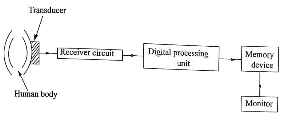
- The received sound signals are converted into digital signal echoes are different and stored in the computer.
- The computer screen displays the image of the structure. The above procedure is known as sonogram or ultrasound scan.

Fig 1.1. Block diagram of sonogram
Working
- The sonography system consist of ultrasonic signal generator, transducers and A, B or C scan display. The block diagram of the experimental setup is shown.
- The transducers are placed nearer the body area of the patient’s skin to be scanned.
- A suitable couplant is used to transmit the sound waves generated by the transducer to pass to the body skin.
- Normally, these couplant are in the form of a gel and are spread over the scanning area. The sound waves are transmitted into body parts and received back by the same transducers.
- The image of the object is obtained in any of the A, B or C scan format.
- The different parts such as head, hands and feet images are also shown in the scan.
Applications
- It is used to examine the interior parts of the body more accurately.
- To monitor the health and development of foetus.
- It is used to confirm pregnancy, ensure that foetus is developing normally and to indicate delivery dates more accurately.
- It is used to examine the birth defects or other parental problems of fetus.
- The problems such as gallstones or liver diseases are examined in the abdomen.
- It is used to identify heart diseases.
Advantages
- It is a non-invariance and non-radiation technique
- It is a harmless technique and does not produce any hazards like bleeding or infections or chemical reactions.
Disadvantages
- It requires a skilled person known as monographer to do the scanning
- It requires a skilled person to analyse the image of the scanned parts.
Monitoring stages
It is used to monitor the different stages of pregnancy.
i) 4 – 5 weeks – Pregnancy visible
ii) 5 – 6 weeks – Fetal visible
iii) 7 weeks – Heart beat visible
At about nine weeks, the baby’s crown – rump length is easily measurable.
| Read More Topics |
| Poisson’s ratio |
| Hooke’s law |
| Newton’s law of cooling |


