Transmission electron microscope utilise an electron beam which passes through and interacts with an ultra-thin sample to produce an image, which is then magnified and focused on an imaging device (a fluorescent screen), on a photographic film layer, or is detected with a sensor (a CCD camera). Max Kroll and Ernst Ruska designed the first TEM in 1931; designed the first TEM with resolution power greater than that of light in 1933; and the first commercial TEM in 1939.
TEM relies on the same basic principle as the light microscope; however it has a greater resolution which allows studying the ultrastructure of organelles, viruses, and. macromolecules. Specially prepared samples can also be observed using TEM. Combination of light microscope and TEM is often used to complement a research. project.
The basic construction of TEM (figure) is discussed below:
Electron Source (Gun) : This forms the foremost and basic part of a TEM. The electron gun consists of a V-shaped filament of LaB6 or W (tungsten), wreathed with Wehnelt electrode (or cap).
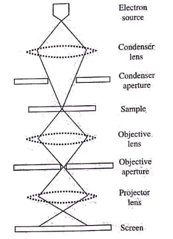
Condenser Lens : The electron stream from the electron gun is focused using the condenser lenses on a small, thin, coherent beam. The first condenser lens determines the spot size (the general size range of the final spot striking the sample), and the second lens changes the spot size on the sample.
Condenser Aperture : This is a thin metal disk or strip having a small circular hole. Aperture is used to restrict the electron beams and filter out the unwanted scattered electrons before forming an image.
Sample : The electron beam after passing the aperture strikes the sample to undergo electron-sample interaction in three different ways, i,e., un-scattered electrons (transmitted beam), elastically scattered electrons'(diffracted-beam), and inelastically scattered electrons.
Objective Lens : This is used to focus the electron transmitted from the sample into an image.
Objective Aperture : This is used to enhance the contrast by blocking out highangle diffracted electrons.
Selected Aperture : The user examines the periodic diffraction of electron by ordered arrangements of atoms in the sample using this aperture.
Projector Lens : This lens is used to expand the beam on the phosphor screen.
Screen : TEM uses a phosphor screen made up of fine (10−100μm) particulate zinc sulphide for direct observation by the user.
Image Pattern : The image on striking the phosphor screen generates a light which allows the user to view the image. The darker areas of image represent those sample areas which transmit fewer electrons; while the lighter areas of image represent those sample areas which transmit more electrons.
In Transmission Electron Microscope (TEM), an electron beam from an electron gun is passed through a series of electromagnetic lenses, which scatter and transmit the beam through the sample and then through the objective lens which magnifies the sample image. The image is then further magnified by the projection lens and projected on the fluorescent screen or photographic film. This converts the electron image into a visible form.
Very thin sections of sample are examined, since the penetration power of electron beam through the solid matter is low. The scattering degree of electrons by the sample is related to the number and mass of atoms lying in the electron path. The contrast of biological materials can be enhanced by staining with salts of heavy metals (like uranium or tungsten) because their constituents are mostly of low mass and the contrast of these materials is also weak.
The heavy metals used for contrasting are either fixed on the sample (positive staining) or they increase the opacity of the surrounding area (negative staining).
Electrons and the sample atoms interact strongly by elastic and inelastic scattering. The sample should be very thin (typically of the order of the 5−100 nm for 100keV electrons), depending on the density and elemental composition of the object and the resolution desired. Special sample preparation techniques are required; e.g., electropolishing and ion-beam etching in materials science, ultramicrotomy of stained and embedded tissues, or cryofixation in biosciences.
Electrons from the electron gun are emitted by thermionic, Schottky, or field emission. The latter is used when high gun brightness and coherence are required. A condenserlens system having three or four stages allows the user to change the illumination aperture and the area of illuminated sample.
The electron intensity distribution behind the sample is imaged on a fluorescent screen using a three to eight lens system. The image on the screen is recorded either by directly subjecting the photographic emulsion or an image plate within a vacuum chamber, or digitally to a CCD camera via fluorescent screen coupled with a fibre-optic plate.
Applications
- It is ideally fised in different fields like life sciences, nanotechnology, medical, biological and material research, forensic analysis, gemology, metallurgy, industry, and education.
- It provides topographical, morphological, compositional, and crystalline information of the sample.
- The images it produces allow the researchers to view the samples on a molecular level, thus making possible to study the ultrastructure and texture of sample.
- It is used for semiconductor analysis and manufacturing of computer and silicon chips.
- It is used in technology companies for identifying flaws, fractures and damages in micro-sized objects, so that with the data obtained the problems can be overcome and/or a more durable and efficient product can be formed.
Advantages
- It has the most powerful magnification (potentially over 1 million times or more).
- It has a wide range of applications and can be utilised in various different scientific, educational, and industrial fields.
- It provides information on element and compound structure.
- It produces high quality and detailed images.
- It provides information on surface features, shape, size, and structure.
- Its operation is easy with proper training.
Disadvantages
- It is large and expensive.
- Sample preparation is a tedious task.
- Potential artefacts from sample preparation.
- Its operation and analysis requires special training.
- Only those samples which are electron transparent can withstand inside the vacuum chamber, and are small enough to fit in the chamber can be used.
- It requires special housing and maintenance.
- It produces black and white images.
[sc_fs_faq html=”true” headline=”h2″ img=”” question=”What is Microscopy?” img_alt=”” css_class=””] Microscopy is the scientific practice of using microscopes to observe and study objects and samples that are too small to be seen with the naked eye. Microscopy allows scientists to visualize the microscopic structures, organisms, and materials that make up our world. [/sc_fs_faq]
| Read More Topics |
| Scanning electron microscopy (SEM) |
| Quantitative measurement of bacterial growth |
| Single cell isolation methods |
| Cultivation of bacteria (Aerobes and anaerobes) |