The physiological barriers are discussed below:
Blood Capillary Membrane: Drug passes the capillary membrane through passive diffusion and hydrostatic pressure. By passive diffusion, drug molecules travel across the region of high concentration to low concentration; it can be described by Fick’s law of diffusion.
![]()
D = Drug diffusion coefficient in the membrane.
K = Lipid/water partition coefficient.
A = Membrane surface area.
CP = Plasma drug concentration.
Ct = Tissue drug concentration.
h = Membrane thickness.
The negative sign indicates drug movement from inside the blood capillary into the tissues.
2) Simple Capillary Endothelial Barrier: Capillaries supply blood to most of the tissues. Capillary endothelium allows passage of drugs (ionised or unionised of molecular size <600 Daltons) into the interstitial fluid. Only drugs bound to the blood components are restricted because of the large molecular size of the complex.
3) Cell Membrane: Drug present in ECF is transported by passive diffusion into the cell. Factors influencing the penetration of drugs into cells are same as those observed in the gastrointestinal absorption of drugs. For the transport of drugs, cell membrane acts like a lipid barrier. Permeability of drugs through cell membrane checks the entry into the cell. The physicochemical properties that influence permeation of drugs across such a barrier are illustrated in figure (A).

Figure (A) Cell Membrane Barrier and Drug Diffusion across it
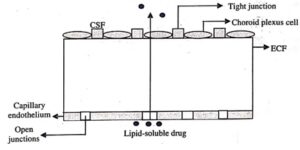
4) Blood-Brain Barrier (BBB): Permeability of capillaries present in the brain and spinal cord is different from that of the capillaries of rest of the body. Endothelial cells of capillaries are covered by a layer of glial cells that have tight intercellular junctions providing thicker lipid barrier. This layer of glial cells reduces diffusion and penetration of water-soluble and polar drugs into the brain and spinal cord. Figure (B) represents this lipid barrier (BBB).

Figure (B) Transport of Lipid-Soluble Drug across BBB
Normally, only lipid-soluble drugs can penetrate the interstitial fluid of the brain and spinal cord, while water-soluble compounds need specific carriers to cross the endothelial lining. In diffusion process, many transport mechanisms are involved. Degree of ionisation in plasma and drug lipid solubility determines the penetration rate of a drug into the brain. Highly lipid-soluble drugs (e.g., thiopental) cross BBB immediately, and reach the brain from plasma. Polar drugs (e.g., barbital) penetrate the CNS slowly. Weak organic acids (e.g., penicillin G having pKa 2.6 ) are found in completely ionised form in plasma, but penetrate the brain at a low rate due to poor lipid solubility.
Approaches to Promote Crossing the BBB
- Permeation enhancers (e.g., Dimethyl Sulfoxide or DMSO) are used for increasing penetration.
- Mannitol infused in internal carotid artery result in osmotic disruption of BBB.
- Carriers (e.g., dihydropyridine redox system) are used to transport drug into brain.
- Blood-Cerebrospinal Fluid Barrier: Cerebrospinal fluid forms in choroid plexus, found in the roof of the fourth ventricle and projects between the cerebellum and pons on the lower brain stem. At anterior brain stem in the roof of the diencephalon, two extensive folds of choroid plexus originate and extend through inter-ventricular foramina. Floors of the lateral ventricles are covered by folds of the choroid plexus.

As the CSF is almost devoid of protein, the CSF concentration of lipidsoluble drugs represents the free drug concentration in plasma. Concentration of drugs is greater in brain tissues as compared to the CSF, e.g., in epileptic patients concentration of phenytoin is 6 times higher in the temporal lobe than in the CSF.
- Placental Barrier: Maternal and the foetal blood vessels are separated by placental barrier, which is made up of endothelium and many layers of tissue of foetal trophoblast basement membrane. Figure (D) shows the blood flow in maternal and foetal blood vessels. Placental barrier is less effective than BBB because drugs having molecular weight less than 1000 Daltons and with moderate to high lipid solubility (e.g., ethanol, sulphonamides, barbiturates, gaseous anaesthetics, steroids, narcotic analgesics, anticonvulsants, and antibiotics) can cross the placental barrier through simple diffusion. Immunoglobulins are transferred by endocytosis, whereas, nutrients essential for foetal growth are transported by carrier-mediated process.

- Blood-Testes Barrier: A layer is formed by the extensions of sustentacular cells (sertoli cells) that surround the seminiferous tubule beneath the spermatogonia. To maintain stable conditions, diffusion from interstitial fluid to the seminiferous tubule is prevented by the tight junctions present between the sustentacular cells.
| Read More Topics |
| Tissue permeability of drugs |
| Introduction to medicinal chemistry |
| Verification of newton’s law of cooling |


