Scanning Electron Microscope (SEM) is an improved model of an electron microscope. SEM is used to study the three dimensional image of the specimen.
Principle
When the accelerated primary electron strikes the sample, it produces secondary electrons. These secondary electrons are collected by a positive charged electron detector which in turn gives a three-dimensional image of the sample.
Construction
It consists of an electron gun to produce high energy electron beam. A magnetic condensing lens is used to condense the electron beam and a scanning coil is arranged in between magnetic condensing lens and the sample. The electron detector (scintillator) is used to collect the secondary electrons which can be converted into electrical signal. This signal is fed into CRO through video amplifier as shown in Fig.

Fig.1.1 Scanning electron microscope
Working
- Stream of electrons are produced by the electron gun and these primary electrons are accelerated by the grid and anode.
- These accelerated primary electrons are made to incident on the sample through condensing lenses and scanning coil.
- These high speed primary electrons on falling over the sample produces low energy secondary electrons.
- The collections of secondary electrons are very difficult because of their low energy.
- Therefore, to collect these secondary electrons, a very high positive voltage is applied to the collector.
- These collected electrons produces scintillation on the photomultiplier tube (detector) and are converted into electrical signals. These signals are then amplified by the video amplifier and are fed to the CRO.
- By similar procedure the electron beam scans the sample from left to right and again from left to right. It is similar to reading a book (Fig .1.2), and the whole picture of the sample is obtained in the CRO screen.
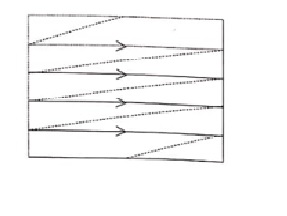 Fig.1.2
Advantages
- It can be used to examine specimens of large thickness.
- It has large depth of focus.
- It can be used to get a three dimensional image of the object.
- Since the image can be directly viewed in the screen, structural details can be resolved in a precise manner.
- The magnification may be up to 3,00,000 times greater than that of the size of the object.
Disadvantage
The resolution of the image is limited to about 10 – 20 nm hence its resolution is very poor.
Applications
- It is used to examine the structure of very large specimens in a three dimensional view.
- It is used to study the microscopic features of the specimen.
- It is used to analyse the IC failure.
- It is used to examine the surface contamination.
- It is used to evaluate the thin coating.
| Read More Topics |
| G.P Thomson Experiment |
| Physical significance of the wave function |
| De-Broglie’s concept of matter |